Dalai's Hospital adds Pet/CT scanner to its Diagnostic Arsenal
Unit is the first one of its kind in the state
Unit is the first one of its kind in the state
Doctor Dalai's Hospital continues its leadership in imaging by adding dedicated PET/CT to its arsenal of diagnostic tools. Dalai's Hospital was the first hospital in the state to introduce positron imaging, initially with coincidence scanning in 1998. Dedicated PET imaging was launched in March 2001 and we now offers the state’s first fixed-site PET/CT. The new scanner combines PET and CT scanner technology to increase diagnostic confidence and improve patient management with faster scan times and higher-quality images.
“At Dalai's Hospital, we are always looking for ways to improve patient outcomes,” said the Hospital's Chief Operating Officer. “We’re glad to continue to lead in this diagnostic area by providing another much needed tool which gives physicians more detailed information to help patients. For us, its part of providing the quality healthcare to our community that Dalai's Hospital is known for.”
Positron Emission Tomography (PET) works by creating images of the biological functions of the body to reveal disease states. Prior to the exam, the patient receives an injection of a tiny amount of radioactive tracer, which emit signals as they travel through the body. Most scans utilize a positron-emitting form of glucose, which allows mapping of metabolism. PET measures the degree of sugar uptake, taking advantage of the fact that cancers use more glucose than normal tissues. The Computed Tomography (CT) aspect of the scanner improves the quality of the PET scan and adds an anatomical basis for localizing where the sugar or glucose uptake has occurred
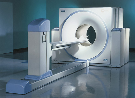
This new class of PET/CT scanner, Siemens Biograph 16, is the first to utilize the proprietary lutetium oxyortho-silicate (LSO) crystals plus true 3D acquisition to allow faster acquisitions of images while improving image quality. No other manufacturer offers this critical technology. For patients, this means more comfort and confidence in the treatment of their cancer and other disease states.
Physicians believe that the improved images produced by the PET/CT could reduce the number of invasive procedures required during follow-up care, including biopsies and even unnecessary operations. “The quality of the images, and the added anatomic dimension, gives us greater ability to find and monitor disease,” said Dr. Dalai, radiologist and director of nuclear medicine at Dalai's Hospital.
Dr. Dalai added, “In fact, images produced by the new scanners are so precise that in some cases we have found malignancy that probably would not have been detected with any other technique. The addition of CT to the PET scan also helps us differentiate normal from abnormal tissues to a much greater degree than we could before.” Dr. Dalai is confident that the new combined PET/CT scanners will play a key role in continuing to improve patient care.
In oncology, PET/CT provides for early diagnosis, more accurate tumor detection and precise localization, improved biopsy sampling, and better assessment of patient responses to chemotherapy and radiation therapy. PET/CT also is used by cardiologists to detect certain types of heart disease and by neurologists for assessing disorders such as Alzheimer’s disease.
No comments :
Post a Comment